Beranda
/ Illustrate A Plant Cell Under Electron Microscope - Illustrate Plant Cells As Seen Under An Electron Microscope Brainly In / Get free solutions to all questions from chapter the fundamental unit of life.
Illustrate A Plant Cell Under Electron Microscope - Illustrate Plant Cells As Seen Under An Electron Microscope Brainly In / Get free solutions to all questions from chapter the fundamental unit of life.
Illustrate A Plant Cell Under Electron Microscope - Illustrate Plant Cells As Seen Under An Electron Microscope Brainly In / Get free solutions to all questions from chapter the fundamental unit of life.. For one thing damage and breaks in structure that are missed in the thick optical sections become glaringly evident even under the optical microscope in the 30 to 100 times thinner sections needed for electron microscopy. Photosynthetic cells of the leaf of elodea. Get free solutions to all questions from chapter the fundamental unit of life. A plant cell as seen under electron microscope major differences between a plant cell and on animal cell are (i) presence of chloroplast in plant cell. Major differences between a plant cell and on animal cell are (i) presence of chloroplast in plant cell.
Organelle location, size, shape and position Here's a diagram of a plant cell: Click here👆to get an answer to your question ️ illustrate only a plant cell as seen under electron microscope. Plant cell as shown above. Get free solutions to all questions from chapter the fundamental unit of life.
The Plant Cell from www.biology-pages.info Labelled diagrams typical animal plant cells stock vector 222613513. A plant cell as seen under electron microscope major differences between a plant cell and on animal cell are (i) presence of chloroplast in plant cell. (a) based on the diagram, state whether it represents an animal cell or plant cell (b) give two reasons for your answer in (a) above (c) why is the palisade layer a tissue? Here's a diagram of a plant cell: Describe how turgor pressure builds up. Illustrate only a plant cell as seen under electron microscope. Click here👆to get an answer to your question ️ illustrate only a plant cell as seen under electron microscope. The cell as seen under the electron microscope.
Since the development of the electron microscope a large number of structures have been identified within cells;
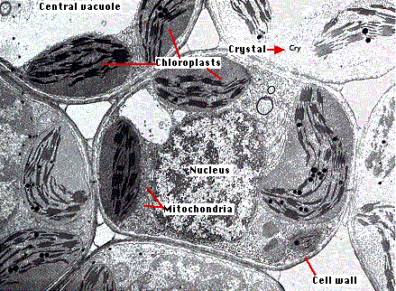
The diagram is very clear, and labeled; Major differences between a plant cell and on animal cell are (i) presence of chloroplast in plant cell. Get free solutions to all questions from chapter the fundamental unit of life. How is it different from animal cell? The electron microscope is more powerful than the light microscope. Plant cell diagram under electron microscope. The electron beam is absorbed or deflected by the heavy metal stains and shadows are cast onto film or a phosphorescent plate (image is a shadow) at the bottom of the column. Organelle location, size, shape and position Illustrate only a plant cell as seen under electron microscope how. The diagram below represents a cell as seen under an electron microscope. How is it different from animal cell? Though we cannot see everything through the light microscope, some important organelles are visible and we can begin to see some structural differences. The electron micrograph displayed below illustrates many of the plant cell characteristics discussed the cell wall, large central vacuole and chloroplasts are clearly visible also visible is the clearly defined nucleus containing chromatin nucleus chromatin the vacuole in this mature plant cell from a leaf is large, and occupies about 80% of
Cell study with a light microscope. It uses a beam of electrons to illuminate the specimen instead of light as in the case of light microscope. Here's a diagram of a plant cell: State two ways in which osmosis is significant to. Those structures with a specialised function are termed organelles.
Botany Online Cells And Tissues What Is Viewed In A Microscope from www1.biologie.uni-hamburg.de Illustrate only a plant cell as seen under electron microscope how. Here's a diagram of a plant cell: Image:plant cell seen under light microscope the cell as seen under the electron microscope. It also has a very high. Made using microsoft sp4 & moviemaker The diagram is very clear, and labeled; Structure of nucleolus under light and electron microscope a. Illustrate only a plant cell as seen under electron microscope.
The cell as seen under the electron microscope.
The plant cell back to menu or next or previous. The electron beam is absorbed or deflected by the heavy metal stains and shadows are cast onto film or a phosphorescent plate (image is a shadow) at the bottom of the column. Illustrate only a plant cell as seen under electron microscope. A typical animal cell (as seen in an electron microscope) medical images for powerpoint 1. How is it different from animal cell? Since the development of the electron microscope a large number of structures have been identified within cells; Once slides have been prepared they can be examined under a microscope. The diagram is very clear, and labeled; How is it different from animal cell? The diagram is very clear, and labeled; Electron column in the microscope. Labelled diagrams typical animal plant cells stock vector 222613513. How is it different from animal cell?
For one thing damage and breaks in structure that are missed in the thick optical sections become glaringly evident even under the optical microscope in the 30 to 100 times thinner sections needed for electron microscopy. Labelled diagram of a plant cell under microscope posted on march 18 2011 by admin onion cells stained with methylene blue look at the images of onion cells as they would be seen under a microscope draw each magnification label appear high picture plant and animal cell diagrams. The diagram is very clear, and labeled; Organelle location, size, shape and position The electron microscope is more powerful than the light microscope.
Illustrate Only A Plant Cell As Seen Under Electron Microscope How Is It Different From Animal Cell from cdn.entrance360.com It also has a very high. Electron microscope can magnify an object up to 500,000 times. The diagram is very clear, and labeled; Labelled diagram of a plant cell under microscope posted on march 18 2011 by admin onion cells stained with methylene blue look at the images of onion cells as they would be seen under a microscope draw each magnification label appear high picture plant and animal cell diagrams. 2 see answers kasi kasi Major differences between a plant cell and on animal cell are (i) presence of chloroplast in plant cell. Illustrate only a plant cell as seen under electron microscope how. Electron microscope can magnify an object up to 500, 000 times.
A plant cell as seen under electron microscope major differences between a plant cell and on animal cell are (i) presence of chloroplast in plant cell.
In truth, there are still features of plant and anim. Though we cannot see everything through the light microscope, some important organelles are visible and we can begin to see some structural differences. Plant cell diagram under electron microscope. The electron beam is absorbed or deflected by the heavy metal stains and shadows are cast onto film or a phosphorescent plate (image is a shadow) at the bottom of the column. Once slides have been prepared they can be examined under a microscope. A typical animal cell (as seen in an electron microscope) medical images for powerpoint 1. Photo album by darcy plant and animal cells under the microscope. Illustrate only a plant cell as seen under electron microscope. A plant cell as seen under electron microscope major differences between a plant cell and on animal cell are (i) presence of chloroplast in plant cell. Made using microsoft sp4 & moviemaker The diagram is very clear, and labeled; But at the same time it is interpretive. How is it different from animal cell?
Made using microsoft sp4 & moviemaker plant cell under electron microscope. Click here👆to get an answer to your question ️ illustrate only a plant cell as seen under electron microscope.
Berbagi :
Posting Komentar
untuk "Illustrate A Plant Cell Under Electron Microscope - Illustrate Plant Cells As Seen Under An Electron Microscope Brainly In / Get free solutions to all questions from chapter the fundamental unit of life."


Posting Komentar untuk "Illustrate A Plant Cell Under Electron Microscope - Illustrate Plant Cells As Seen Under An Electron Microscope Brainly In / Get free solutions to all questions from chapter the fundamental unit of life."